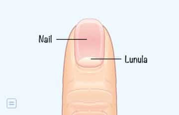
The half-moon shape at the base of your fingernail is known as a lunula. Lunulae cover the bottom of your nail, just above your cuticle.
[ninja_tables id=”73415″]
Lunulae are part of your nail matrix. The matrix refers to the tissue just beneath your nail. It contains nerves, lymph, and blood vessels. It also produces the cells that become the hardened nail plate, which is what you see.
Although everyone has a nail matrix, not everyone will see or have a lunula on each nail. Those who do have a lunula may notice that they vary in appearance across each nail.
Decorate your house with Square Canvas Photo Prints for wall art. Stylish, customizable, and perfect for showcasing your favorite photos.
Read on to learn more about what these lunalae look like, when their appearance or absence could be cause for concern, and when to see your doctor.
An Overview of the Lunala

The lunula, or lunulae (pl.) (derived from a Latin word meaning ‘little moon’), is the crescent-shaped whitish area of the bed of a fingernail or toenail.[mfn]https://en.wikipedia.org/wiki/Lunula_(anatomy)[/mfn]
In humans, it appears by week 14 of gestation and has a primary structural role in defining the free edge of the distal nail plate (the part of the nail that grows outward).
The video below expatriate on the lunala’s significance:
The Appearance of the Lunala
It is located at the end of the nail (that is closest to the skin of the finger), but it still lies under the nail. It is not actually white but only appears when it is seen through the nail.
Outlining the nail matrix, the lunula is a very delicate part of the nail structure. If one damages the lunula, the nail will be permanently deformed. Even when the totality of the nail is removed, the lunula remains in place and is similar in appearance to another smaller fingernail embedded in the nail bed.
In most cases, it is half-moon-shaped and has unique histologic features. Examinations concluded that the lunula is an area of the loose dermis with lesser-developed collagen bundles. It appears whitish because a thickened underlying stratum basale obscures the underlying blood vessels.
The lunula is most noticeable on the thumb; however, not everyone’s lunulae are visible. In some cases, the eponychium may partially or completely cover the lunula.

The Importance of the Lunala
The lunula’s location on the newest part of the nail allows assessments to be made about one’s health. In some cases, its absence may indicate an underlying health problem. Some examples of such problems are malnourishment, anemia, kidney failure, and heart disease.
What does a Healthy Lunulae look like?

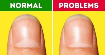
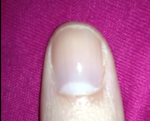
Healthy lunulae are usually a whitish color and take up a small portion of the bottom of your nail. They’re usually most visible on your thumb.
You may notice that they appear smaller on your pointer finger, gradually shrinking in size until you reach your pinkie where they may be barely visible.
What if your Lunulae Change Color?
Sometimes, the appearance of your lunula or overall nail can be a sign of an underlying condition.
Check the table below to know what a lunalae color change indicates;

What Causes Abnormal Coloring of the Lunulae?

Here are some of the most common reasons for abnormal lunulae:
- Tetracycline therapy
Tetracycline medications are antibiotics that are usually used to treat acne and skin infections. Extended use may cause your lunulae to turn yellow.
- Diabetes
Pale blue lunulae may be a sign of undiagnosed or uncontrolled diabetes. This is a chronic, lifelong condition that affects the body’s ability to control blood sugar.
- Excessive fluoride ingestion
Taking in too much fluoride, like that found in toothpaste, can turn the lunulae brown or black.
- Silver poisoning
Blue-gray lunulae may be a sign of silver poisoning.
- Yellow nail syndrome
This condition typically produces thick, slow-growing nails. The middle of your nail may begin to rise, causing the lunulae to disappear completely. Your entire nail will take on a yellow appearance.
It isn’t clear what causes this syndrome, but it may be tied to:
- chronic sinusitis
- pleural effusion
- recurrent pneumonia
- lymphedema
- rheumatoid arthritis
- immunodeficiency disorders
- Terry’s nails
This condition causes the bulk of your nail to appear white, completely erasing the appearance of the lunula. It’s characterized by a pink or red band of separation near the arc of your nails. Although it can happen on one finger only, it usually affects all fingers.
In older adults, this condition is usually a natural sign of aging.
In some cases, it may be a sign of:
- diabetes
- liver disease
- kidney failure
- congestive heart failure
- Wilson’s disease
This is a rare inherited disorder that occurs when too much copper accumulates in your organs. It’s known to cause blue lunulae.
- Severe renal disease
The portion of your nail containing the lunula may turn white, sometimes creating a nail that’s half-brown and half-white. This is sometimes called half-and-half nails and may be a sign of renal disease.[mfn]healthline[/mfn]
- Chronic renal failure
People who experience chronic renal failure may produce more melanin, which can cause your nail bed to turn brown.
- Heart failure
If your lunula turns red, it may be a signal of heart failure.
What Does it Mean if your Lunulae are Small or Missing?
Small or missing lunulae usually aren’t usually a cause for concern. They’re usually just hidden underneath the cuticle or skin at the base of your finger.
In some cases, missing lunulae may be a result of trauma or a sign of:
- anemia
- malnutrition
- depression
If you’re experiencing other unusual symptoms, such as fatigue or overall weakness, make an appointment to see your doctor. They can perform a physical exam to help diagnose the cause of your symptoms and advise you on the next steps.
What if your Lunulae are Large or Misshaped?

Researchers don’t know what causes the lunula to take up a significant portion of the nail.
Some reports suggest that lunulae may signal issues with the cardiovascular system, heartbeat disruption, and low blood pressure.
Unscientific theories claim that large lunulae may be common in athletes and people who engage in lots of physical activity.[mfn]Guglielmi, Giuseppe; Kuijk, Cornelis Van; Genant, Harry K (2001). Fundamentals of hand and wrist imaging. ISBN 978-3-540-67854-0.[/mfn] This may be due to the bodily stress associated with high-impact activities, but there is currently no research to back up these claims.
When to See the Doctor
Discolored or missing lunulae usually aren’t usually a cause for concern. But if you notice changes in your nail appearance and are experiencing other unusual symptoms, make an appointment to see your doctor.
You should seek immediate medication attention if your hands and feet are also turning blue. This could be a sign of cyanosis, a condition that results from poor circulation or inadequate oxygenation of your blood.
Your doctor can assess your symptoms and advise you on treatment options. Treating the underlying condition will usually restore your nail appearance and improve your overall well-being.
Conclusion
The lunula is the visible portion of the distal nail matrix that extends beyond the proximal nail fold. It is white, half-moon-shaped, appears by week 14 of gestation, and has unique histologic features. The lunula has a primary structural role in defining the free edge of the distal nail plate.
Feel free to contact us at [email protected] if you have further questions to ask or if there’s anything you want to contribute or correct to this article.
You can always check our FAQs section below to know more about the lunala.
[bwla_faq faq_topics=”frequently-asked-questions-about-the-lunala” sbox=”1″ paginate=”1″ pag_limit=”5″ list=”1″ /]


