An Overview of Left Axis Deviation
Quick Facts About Left Axis Deviation (LAD)
| A | B |
|---|---|
| Definition | LAD is a condition where the mean electrical axis of ventricular contraction lies between −30° and −90° |
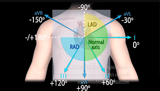
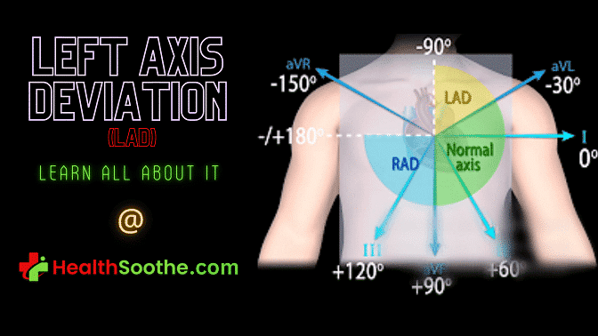
| Electrical Axis Classification | Normal axis (−30° to +90°), Right axis deviation (>+90°), Extreme axis deviation (−90° to 180°) |
| ECG Indication | Positive QRS complex in lead I and negative in leads aVF and II |
| Causes | Various conditions can cause LAD, including: Left anterior fascicular block (electrical conduction block); Left ventricular hypertrophy (enlarged heart muscle); Pre-excitation syndromes (abnormal electrical pathways); Congenital heart defects; Mechanical shifts of the heart (pregnancy, enlarged organs) |
| Symptoms | Symptoms depend on the underlying cause |
| Treatment | Treatment is based on the underlying condition causing LAD |
| Significance | LAD itself is not a diagnosis but can indicate underlying heart problems. Further evaluation by a doctor is usually recommended |
| Special Cases | LAD can be a normal finding in athletes. However, if accompanied by other abnormalities, it might warrant investigation |
Left axis deviation (LAD) is a condition in electrocardiography in which the average electrical axis of the ventricular contraction of the heart rests in a frontal plane direction between 30° and 90°. This is mirrored by a positive QRS complex in lead I and a negative complex in leads aVF & II.
LAD can be caused by a number of factors. Normal variation, pre-excitation syndrome, conduction defects, inferior wall myocardial infarction, congenital heart disease, ventricular ectopic rhythms, emphysema, mechanical shift, high potassium levels, paced rhythm, and thickened left ventricle are just a few of the causes.
The underlying cause determines the symptoms and treatment for left axis deviation.
Left Axis Deviation (LAD) – Defining it

Left axis deviation (LAD) is a condition in electrocardiography in which the average electrical axis of the ventricular contraction of the heart rests in a frontal plane direction between 30° and 90°1https://en.wikipedia.org/wiki/Left_axis_deviation. This is mirrored by a positive QRS complex in lead I and a negative complex in leads aVF & II.
In electrocardiography, the cardiac axis is the total of the depolarization vectors created by each cardiac myocyte. To understand the cardiac axis, one must first discover the connection between both the QRS axis and the ECG limb leads.
Because the left ventricle makes up the majority of the heart muscles, a typical cardiac axis is downward but also slightly to the left. QRS is somewhere between -30° & +90° on a normal axis.
In contrast, LAD is defined as a QRS axis between 30° and 90°, right axis deviation (RAD) is defined as a QRS axis higher than +90°, and extreme axis deviation (EAD) is defined as a QRS axis between -90° to 180°.
Watch the video below to know more on left axis deviation:
Calculating The Left Axis Deviation of the Heart
Knowing the electrical axis may assist guide the differential diagnosis and offer insight into underlying illness conditions2Jenkins, Dean (1996). "The electrical axis at a glance". www.ecglibrary.com. Retrieved 2022-10-25.. The ECG axis may be determined in a variety of ways. The quadrant technique, which looks at lead aVF, and Lead I is the simplest.
First, analyze the QRS complex for both leads I and avF to determine if it is +ve (height of R wave > height of S wave), equiphasic (R wave = height of S wave), or negative (R wave height of S wave). When lead I is +ve while lead aVF is -ve, this might be a case of LAD.
Check QRS in lead II to identify a real LAD. If the QRS complex in lead II is positive, this indicates a normal axis. If, in contrast, the QRS complex in lead II is negative, this indicates a LAD. The Isoelectric lead is another technique of measuring LAD that allows for a more exact calculation of the axis of the QRS.
Causes of Left Axis Deviation
LAD may be caused by a number of factors. Inferior wall myocardial infarction, left ventricular hypertrophy3"Left ventricular hypertrophy - Diagnosis and treatment - Mayo Clinic". www.mayoclinic.org. Retrieved 2022-10-25., ventricular ectopic arrhythmias, congenital cardiac disease, preexcitation syndrome, pacemaker-generated paced rhythm, conduction abnormalities, mechanical shift, emphysema, normal variation, and hyperkalemia are all examples of these.
The normal variation that causes LAD is a physiologic alteration that occurs with age. LAD on the ECG may be caused by conduction problems like a block of the left anterior fascicular branch or left bundle branch block.
LAD on ECG may be caused by pre-excitation syndrome in addition to congenital cardiac abnormalities like atrial septal defect and endocardial cushion deficiencies. An abdominal tumor, Wolff-Parkinson White syndrome, an inferior MI, an enlarged liver or spleen, expiration or a higher diaphragm from pregnancy, or ascites (fluid buildup in the abdomen) are all mechanical alterations that induce LAD.
In athletes, LAD is a borderline trait that, when paired with some other borderline feature like the block of the right bundle branch, necessitates additional evaluation because of the increased likelihood of sudden cardiac death.
Symptoms and Signs of Left Axis Deviation
The symptoms of left axis deviation are determined by the underlying reason. For example, if LAD is caused by left ventricular hypertrophy4"What is Left Ventricular Hypertrophy (LVH)?". www.heart.org. Retrieved 2022-10-25., symptoms may include palpitations, weariness, dizziness, chest discomfort (particularly with exercise), shortness of breath, or fainting.
If a conduction defect, like left bundle branch block, causes LAD, there may be no symptoms except if the conduction malfunction is induced by heart failure, which may lead to heart failure symptoms such as exhaustion or shortness of breath.
Treatment of Left Axis Deviation
Although the left axis deviation may not need therapy in and of itself, the root cause can be addressed. If LAD is caused by left ventricular hypertrophy, therapy is determined by the underlying etiology of the enlargement.
If high blood pressure is the cause of LVH, medications such as diuretics, angiotensin receptor blockers (ARBs), beta-blockers, angiotensin-converting enzyme inhibitors (ACE inhibitors), and calcium channel blockers are used to lower blood pressure and prevent further enlargement of the left ventricle.
If LVH is caused by valvular abnormalities like aortic valve stenosis, the valve must be surgically repaired or replaced.
Is the Left Axis Deviation of the Heart Life Threatening? – Is Left Axis Deviation ECG Dangerous or Can LAD Cause Death?
Although not a dangerous finding in and of itself, axis deviation may be an indication of a serious underlying condition. A careful history to elicit acute cardiac injury is therefore of utmost importance.
In conclusion, among patients with left bundle branch block, those with left axis deviation have a greater incidence of myocardial dysfunction, a more advanced conduction disease, and greater cardiovascular damage which can lead to mortality (if not properly treated immediately) than those with a normal axis.
All right, guys, that is it for now for the left axis deviation of the heart. I hope Healthsoothe answered any questions you had concerning the left axis deviation of the heart.
Feel free to contact us at contact@healthsoothe.com if you have further questions to ask or if there’s anything you want to contribute or correct to this article. And don’t worry, Healthsoothe doesn’t bite.
You can always check our FAQs section below to know more about left axis deviation. And always remember that Healthsoothe is one of the best health sites out there that genuinely cares for you. So, anytime, you need trustworthy answers to any of your health-related questions, come straight to us, and we will solve your problem(s) for you.
Frequently Asked Questions About Left Axis Deviation
What Does Left Axis Deviation Tell You?
Although left axis deviation is often an age-related physiological change. It may indicate the presence of various conditions, such as left ventricular hypertrophy, left anterior fascicular block, inferior wall myocardial infarction, emphysema, and mechanical shift due to elevated diaphragm because of obesity.
Is Left Axis Deviation ECG normal?
The abnormal left axis deviation is one of the most common abnormal ECG findings. Among 67,375 Air Force men without symptoms, Hiss and associates found a frontal plane QRS axis of −30 to −90 degrees in 128 (1.9 percent).
Should I be Worried About Left Axis Deviation?
Although not a dangerous finding in and of itself, axis deviation may be an indication of a serious underlying condition. A careful history to elicit acute cardiac injury is therefore of utmost importance.
Does Left Axis Deviation mean that You Have Heart Disease(s)?
A research was carried out, and the results were that the development of left axis deviation in people of 40-59yr of age, independent of blood pressure is a significant predictor of ischemic heart disease events that are usually manifest 5-10yr after the onset of this electrocardiographic abnormality.
Can Obesity Cause Left Axis Deviation?
The prevalence of left-axis deviation (LAD) (QRS axis of -30 degrees or less) was not higher among those with greater measures of body fatness. There were no significant differences in mean age-adjusted skinfold thickness, height, weight, or chest circumference between those with LAD and those with a normal QRS axis.
Additional resources and citations
- 1https://en.wikipedia.org/wiki/Left_axis_deviation
- 2Jenkins, Dean (1996). "The electrical axis at a glance". www.ecglibrary.com. Retrieved 2022-10-25.
- 3"Left ventricular hypertrophy - Diagnosis and treatment - Mayo Clinic". www.mayoclinic.org. Retrieved 2022-10-25.
- 4"What is Left Ventricular Hypertrophy (LVH)?". www.heart.org. Retrieved 2022-10-25.


