
Medically Reviewed
This article has been reviewed for clinical accuracy.
NECROTIZING ULCERATIVE GINGIVITIS
Necrotizing ulcerative gingivitis (NUG) is a non-contagious anaerobic inflammatory destructive disease of the gingiva which is associated with an overwhelming proliferation of Borrelia Vincenti and fusiform bacteria presenting characteristic signs and symptoms.
TERMINOLOGIES
• Trench mouth
• Vincent’s Stomatitis
• Vincent’s Infection
• Fusospirochetal Gingivitis
• Necrotizing Gingivitis
EPIDEMIOLOGY & PREVALENCE
- Often occurs in groups in an epidemic pattern.
- The prevalence of ANUG appears to have been rather low in the United States and Europe prior to 1914.
- The incidence of ANUG was reported as 0.08% in patients 15 to 19 years old, 0.05% in those 20 to 24 years old, and 0.02% in persons 25 to 29 years old.
- In India, 54% and 58% of the patients in two studies were under age 10.
- Age group affected 18-30 years.
- Not common in children.
- Reported in children from low socio-economic groups in underdeveloped countries.
- In India, 54% and 58% of the patients in two studies were under age 10.
- Age group affected 18-30 years.
- Not common in children.
- Reported in children from low socio-economic groups in underdeveloped countries.
TYPES
According to Pindborg J (1951)-classified into 3 forms:
- Acute
- Subacute
- Chronic (Now considered misidentification of recurrence of the disease).
ETIOLOGY
ROLE OF BACTERIA
- Plaut in 1894 and Vincent in 1896 first proposed this bacterial etiology.
- They both reported that fusiform-spirochete bacteria flora were associated with lesions of NUG.
- Roseburry described a fusospirochetal complex consisting of T. macrodentium, intermediate spirochetes, vibrios, fusiform bacilli, and filamentous organisms in addition to several Borrelia species.
- In India, 54% and 58% of the patients in two studies were under age 10.
- Age group affected 18-30 years.
- Not common in children.
- It has been Reported in children from low socio-economic groups in underdeveloped countries.
- More common in children with Down’s syndrome than in children with mental deficiencies.
LOCAL PREDISPOSING FACTORS
- Pre-existing gingivitis,
- Injury to the gingiva, and smoking.
- Although ANUG may appear in an otherwise disease-free mouth, it most often occurs superimposed on preexisting chronic gingival disease and periodontal pockets.
- Deep periodontal pockets and pericoronal flaps
- Areas of the gingiva traumatized by opposing teeth in malocclusion
SYSTEMIC PREDISPOSING FACTORS
Nutritional Deficiency: This results in marked tissue depletion of key antioxidant nutrients, & impaired acute-phase protein response to infections.
- Other features include,
- Inverted helper/T-cell ratio,
- Histaminemia, hormonal imbalance
- blood & saliva levels of free cortisol
- Defective mucosal integrity.
- Deficient macro & micro nutrients, hence poor prognosis of such infections.
Debilitating Disease: a Systemic disease that impairs immunity cause ANUG.
- A disease that predisposes to ANUG is
- Chronic diseases such as syphilis, cancer
- Severe gastrointestinal disorders (ulcerative colitis)
- Blood dyscrasias (Leukemia and anemias)
- A.I.D.S.
- Nutritional deficiencies resulting from debilitating disease may be an additional predisposing factor.
Psychosomatic Factors
During periods of psychological stress, oral hygiene measures may decrease, nutrition may become inadequate, tobacco smoking may increase and immune function may be suppressed. Stressful life events may lead to an activation of the hypothalamic pituitary adrenal axis, resulting in an elevation of serum and urine corticosteroid levels.
Psychologic Stress, Inadequate Sleep
Corticosteroid & Catecholamine levels
Gingival Microcirculation
Neutrophilic and Lymphocytic activity
Enhances tissue damage.

The specific cause of Necrotizing ulcerative gingivitis has not been established. The prevalent opinion is that it is produced by a complex of bacterial organisms but requires underlying tissue changes to facilitate the pathogenic activity of the bacteria.
CLINICAL COURSE
- Indefinite.
- Left untreated, it may lead to Necrotizing ulcerative Periodontitis with a progressive destruction of the periodontium and denudation of the roots, accompanied by an increase in the severity of toxic systemic complications.
- Horning and Cohen 1995
STAGE 1: Necrosis of tip of the Interdental papilla
STAGE 2: Necrosis of entire papilla
STAGE 3: Necrosis extending to the gingival margin
STAGE 4: Necrosis extending to the Attached gingiva
STAGE 5: Necrosis of Buccal/Labial Mucosa
STAGE 6: Necrosis exposing Alveolar Bone
STAGE 7: Necrosis perforating skin of cheek
- Stage 1=NUG,
- Stage 2 = either NUG or NUP because attachment loss may have occurred,
- Stages 3 and 4 = NUP,
- Stages 5 and 6 = Necrotizing Stomatitis, and
- Stage 7 = Noma.
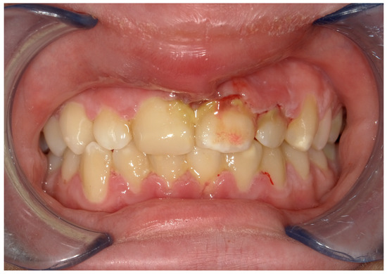
ORAL SIGNS
- Punched out crater-like depressions at the crest of interdental papillae, extending to the marginal gingiva.
- Grayish pseudo-membrane slough covering the craters.
- Spontaneous bleeding on slightest stimulation.
- Fetid odor
- Increased pasty saliva.
- Metallic foul taste.
- Extreme sensitive to touch.
SYSTEMIC SIGNS & SYMPTOMS
- Mild-moderate stages – local lymphadenopathy, slight elevation in temperature.
- Severe cases – high fever, increased pulse rate, loss of appetite.
- Rarely – sequel such as noma.
DIFFERENTIAL DIAGNOSIS
- NUG should be differentiated from other conditions that resemble it in some respects such as:
- Herpetic gingivostomatitis
- Chronic periodontitis
- Desquamative gingivitis
- Streptococcal gingivostomatitis
- Diphtheritic and syphilitic lesions
- Agranulocytosis.
DIAGNOSIS
Diagnosis is based on clinical findings.
A bacterial smear may be used to corroborate the clinical diagnosis, but it is not necessary nor definitive because the bacterial picture is not appreciably different from the other conditions (Gingivitis, periodontal pockets etc).
TREATMENT
- Alleviation of the acute inflammation.
- Treatment of chronic disease either underlying the acute involvement or elsewhere in the oral cavity.
- Alleviation of generalized toxic symptoms such as fever or malaise.
- Correction of systemic conditions that contribute to the initiation or progress of the gingival changes.
- Treatment should follow an orderly sequence and is divided into treatment for
- Non-ambulatory patient: With symptoms of generalized systemic complications.
- Ambulatory patient: With no serious systemic complications
Non-Ambulatory Patients
Day 1:
- Local treatment limited to gently removing the necrotic pseudomembrane with a pellet of cotton saturated with hydrogen peroxide (H2O2)
- Advised bed rest and rinse the mouth every 2 hours with a diluted 3 percent hydrogen peroxide.
- Systemic antibiotics like penicillin or metronidazole can be prescribed.
- Day 2:
- If a condition is improved, proceed to the treatment described for ambulatory patients. If there is no improvement at the end of the 24 hours, a bedside visit should be made. The treatment again includes gently swab the area with hydrogen peroxide, instructions of the previous day are repeated.
- Day 3:
- Most cases, the condition will be improved, start the treatment for ambulatory patients.
Ambulatory Patients
- First visit:
- A clinician should obtain a general impression of the patient’s background, including information regarding recent illness, living conditions, dietary background, type of employment, hours of rest and mental stress.
- Patient’s general appearance should be observed, apparent nutritional status and responsiveness or lassitude, and temperature noted.
- Sub Maxillary and Sub Mental lymph nodes palpated.
- Oral cavity examined for a characteristic lesion of NUG, it’s distribution and a possible involvement of the oropharyngeal region.
- Oral hygiene evaluated, the presence of pericoronal flaps, periodontal pockets, and local irritants determined.
- Patient questioned regarding a history of the acute disease and its onset and duration, recurrence; previous treatment – how long, type of treatment.
- Initial treatment confined to acutely involved areas, which are isolated with cotton rolls and dried.
- Topical anesthesia applied and area gently swabbed with a cotton pellet to remove the pseudo membrane and non-attached surface debris.
- Area cleansed with warm water, superficial calculus is removed.
- Subgingival scaling and curettage contraindicated to prevent extension of infection to deeper tissues and also bacteremia.
- Unless an emergency exists, extractions or periodontal surgery are postponed until the patient has been symptom-free for a period of 4 weeks to minimize the likelihood of exacerbation.
- Patients with moderate or severe NUG and local lymphadenopathy or other systemic symptoms are placed on an antibiotic regimen of Penicillin (500mg 6th hourly);
- Metronidazole 500mg bid 7 days is also effective.
- Antibiotics continued until systemic complications subside.
INSTRUCTIONS AFTER THE FIRST VISIT
- Avoid tobacco, alcohol, and condiments.
- Rinse with a glassful of an equal mixture of 3% H2O2 and warm water every 2 hours and/or twice daily with 0.12% chlorhexidine solution.
- Avoid excessive physical exertion or prolonged exposure to the sun.
- Confine tooth brushing to the removal of surface debris with a bland dentifrice overzealous brushing and the use of dental floss or interdental cleaners will be painful.
- Second visit:
- 1 to 2 days later, the patient’s condition is usually improved; pain diminished or no longer present.
- Gingival margins of the involved areas are erythematous, but without a superficial pseudo membrane.
- Scaling performed if sensitivity permits.
- Instructions as same as above.
- Third visit:
- 1 to 2 days after the second visit, a patient should be essentially symptom free.
- Scaling and root planing repeated.
- Instructed in plaque control procedures.
- H2O2 rinses discontinued; chlorhexidene rinses maintained for 2-3 weeks.
- Fourth visit:
- Oral hygiene instructions are reinforced and thorough scaling and root planing are performed.
- Fifth visit:
- Appointments are fixed for treatment of chronic gingivitis, periodontal pockets and pericoronal flaps, and for the elimination of all local irritants. Patient is placed on maintenance programme.
Gingival Changes With Healing
- Removal of surface pseudo membrane exposes the underlying red, hemorrhagic, craterlike depressions in the gingival.
- In the next stage, the bulk and redness of the crater margins are reduced, but the surface remains shiny.
- Early signs of restoration of normal gingival contour and color follow this.
- Final stage: normal gingival color, consistency, surface texture and contour are restored. Portions of the root exposed by the acute disease are covered by healthy gingiva.
CONCLUSION
- Local procedures are the keystones of the treatment of necrotizing ulcerative gingivitis.
- Inflammation is a local conditioning factor that impairs the nutrition of the gingiva regardless of the systemic nutritional status.
- Local irritants should be eliminated to foster normal metabolic and reparative processes in the gingival.
- Persistent or recurrent Necrotizing ulcerative gingivitis is more likely to be caused by the failure to remove local irritants and by inadequate plaque control than by nutritional deficiency.