
Introduction to the Fossa Ovalis
[ninja_tables id=”73093″]
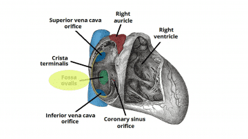
The fossa ovalis is a depressed structure, of varying shapes, located in the inferior aspect of the right interatrial septum [mfn]Malik SB, Kwan D, Shah AB, Hsu JY. The right atrium: gateway to the heart–anatomic and pathologic imaging findings. Radiographics. 2015 Jan-Feb;35(1):14-31. [PubMed][/mfn].
A remnant of an interatrial opening, the foramen ovale, which has a significant role in fetal circulation, the fossa ovalis forms by the fusion of the septum primum and septum secundum [mfn]Joshi SD, Chawre HK, Joshi SS. Morphological study of fossa ovalis and its clinical relevance. Indian Heart J. 2016 Mar-Apr;68(2):147-52. [PMC free article] [PubMed][/mfn].
This article will describe the structure, function, embryology, vascular supply, physiologic variants, surgical considerations, and clinical significance of the fossa ovalis.
The Structure and Function of the Fossa Ovalis

The fossa ovalis is a depressed structure located in the lower portion of the right atrial side of the interatrial septum.
Although the fossa ovalis appears two-dimensional, it is, in fact, a three-dimensional structure – consisting of the septum primum, septum secundum, and the annulus or limbus fossa ovalis[mfn]https://en.wikipedia.org/wiki/Fossa_ovalis_(heart)#:~:text=The%20fossa%20ovalis%20is%20a,foramen%20ovale%20during%20fetal%20development[/mfn] which raises around the perimeter of the fossa ovalis.
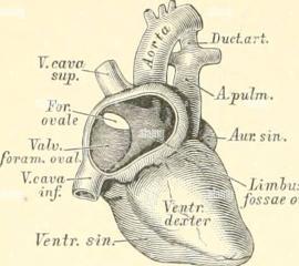
Important neighboring structures are as follows: below and rightward of the fossa ovalis lies the inferior vena cava opening; the location of the coronary sinus is anterior to the fossa ovalis; along the same horizontal plane of the fossa ovalis is the His bundle.
The fossa ovalis forms part of the atrial septum which appears expansive from the right atrial view. However, the true and false atrial septa are clinically important features that need to be distinguished.
The true interatrial septum contains the fossa ovalis and comprises only 20% of the entire septum. It is the only area through which the interatrial septum may be traversed without the risk of cardiac perforation.
In the normal heart, the fossa ovalis serves to prevent blood flow, i.e., shunting of blood, across the interatrial septum.
The Embryology of the Fossa Ovalis

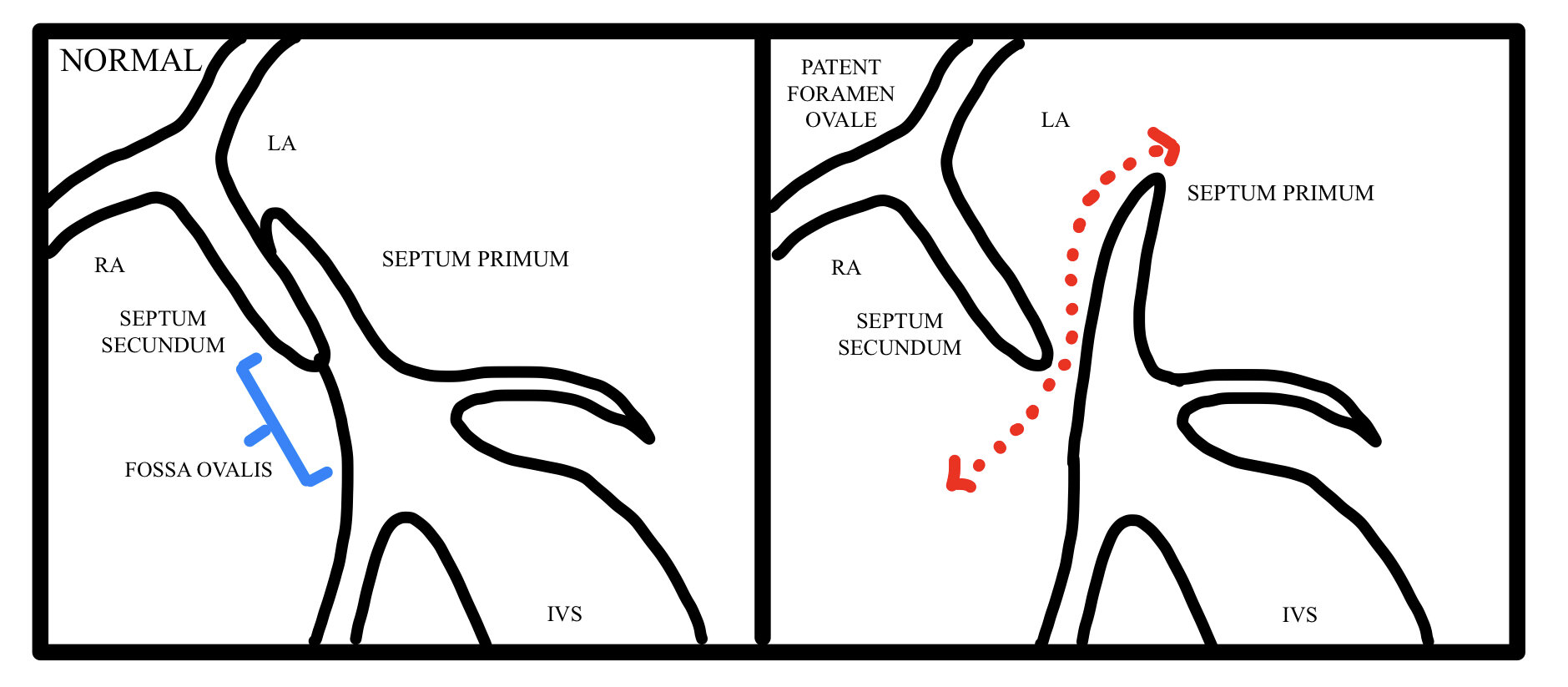
During fetal life, ostia within the juxtaposed septa (septum primum and septum secundum) form a one-way flap valve, the foramen ovale, that allows shunting of blood from the right atrium to the left atrium.
The septum primum which is on the left atrial side forms the floor of the fossa ovalis, whereas the septum secundum derived from an infoldings of the roof of the atrium forms the limbus of the fossa ovalis.
Physiologic increase in the left heart pressure at the time of birth allows the septum primum to create a tight seal with the septum secundum, creating the fossa ovalis.
This fusion of the septum primum with the septum secundum and subsequent formation of the fossa ovalis occurs in 75% of cases, failure of which leads to a patent foramen ovale. Also, the fossa ovalis undergo thinning in utero.
The Blood Supply and Lymphatics of the Fossa Ovalis

The interatrial septum (IAS) receives vascular supply by anastomoses between the left and right branches of the anterior and posterior atrial branches of the right and left coronary arteries; termed Kugel’s artery or arteria anastomotica auricularis magna.
The fossa ovalis together with the middle part of the IAS receives the least vascular supply, additionally, the number and density of these vessels’ networks decrease with age, whereas the anteroinferior portion of the septum receives the densest vascular network.
The Nerves of the Fossa Ovalis
The right atrium has an angiotensinergic innervation and is a source of angiotensin II. The peripheral fibers are mainly non-catecholaminergic afferents and vagal efferents with a small amount being sympathetic.
The Muscles of the Fossa Ovalis
The pectinate muscles or musculi pectinati compose the walls of the atria. They are parallel ridges in the walls of the right atrium. The crista terminalis is a smooth muscular ridge in the superior portion of the right atrium. It divides the musculi pectinati and the right atrial appendage from the smooth surface of the right atrium.
The Physiologic Variants of the Fossa Ovalis
The fossa ovalis may assume various shapes and dimensions [mfn]Rana BS, Shapiro LM, McCarthy KP, Ho SY. Three-dimensional imaging of the atrial septum and patent foramen ovale anatomy: defining the morphological phenotypes of patent foramen ovale. Eur J Echocardiogr. 2010 Dec;11(10):i19-25. [PubMed][/mfn]. It most commonly assumes an oval shape but may be circular or even elliptical.
The dimensions of the fossa ovalis vary among populations as well as by the measuring tools employed but may correlate with the patient’s weight of the heart, age, and body weight.
Significant variations of the fossa ovalis exist, with recesses, slits, aneurysms, and fibrous strands found in certain specimens.
The Surgical Considerations of the Fossa Ovalis
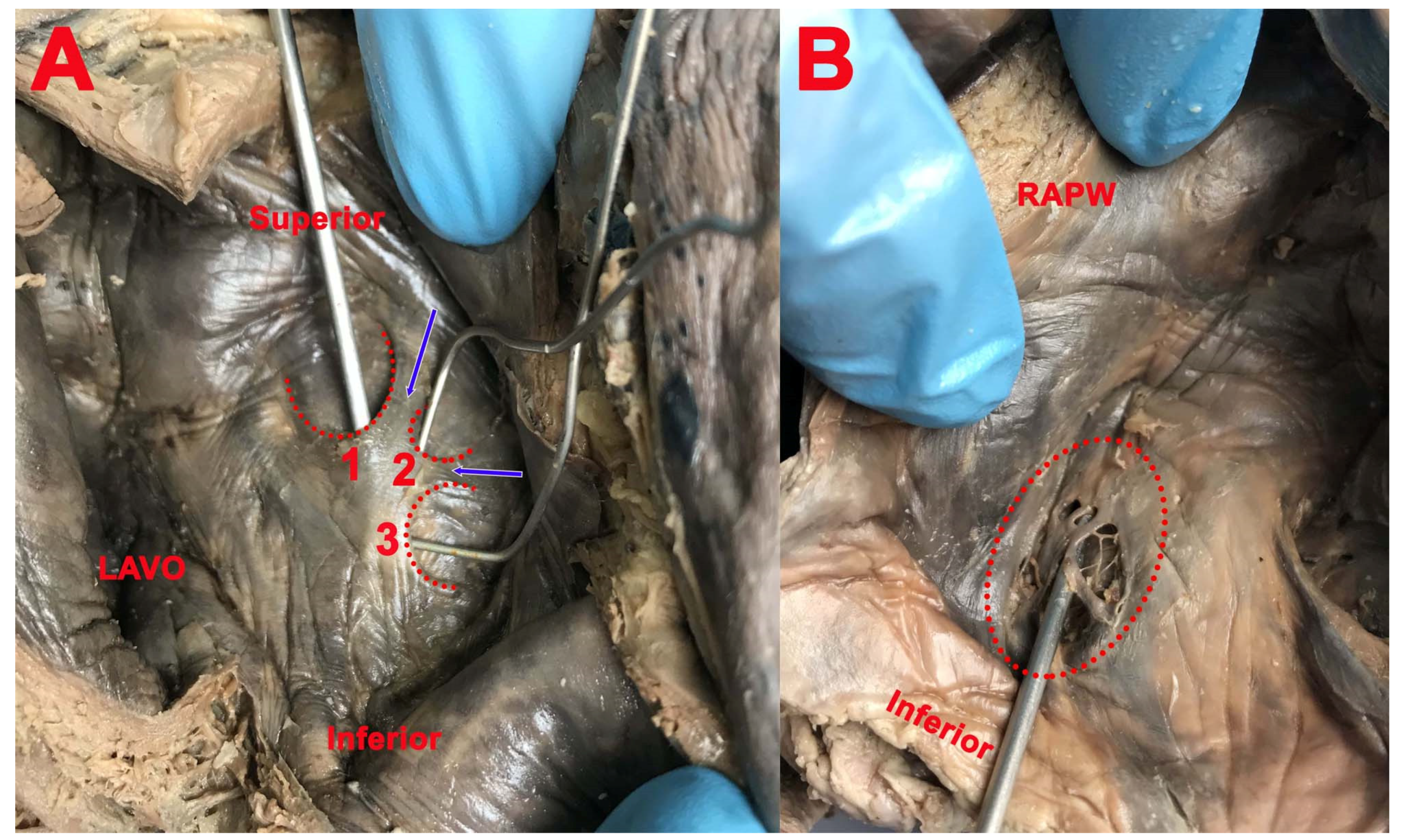
The (limbus) fossa ovalis represents the most direct anatomical landmark for atrial septal puncture for various procedures and may be located using the His bundle electrogram locating method.
The following are significant procedures that require the location of the fossa ovalis for transseptal puncture:
- Patent foramen ovale and atrial septal defect repair
- Right heart catheterization
- Percutaneous balloon valvuloplasty
- Radiofrequency catheter ablation
- Pulmonary vein isolation
- Left atrial appendage closure
- Catheter-based mitral valve repair
- Hemodynamic assessment of the mitral valve
- Paravalvular leak closure
- Alternative access to the left ventricle in the presence of a prosthetic aortic valve
Intracardiac echocardiography (ICE) and fluoroscopy are useful for identifying the fossa ovalis, a critical landmark for electrophysiological procedures.
The Clinical Significance of the Fossa Ovalis
The fossa ovalis may be a frequent site of cardiac pathology. Known pathologies involving the fossa ovalis include:
- Premature closure of the foramen ovale & Patent foramen ovale (PFO)
- Atrial septal defect (ASD)
- Rheumatic heart disease
- Amyloidosis
- Tumors: Prichard’s structures, myxomas, lipomatous hypertrophy of the cardiac interatrial septum
- Fossa ovalis membrane aneurysm (FOMA)
- Cardiac arrhythmias
Patent foramen ovale and atrial septal defect represent functionally similar pathologies; however, they differ in their etiology as well as clinical severity.
ASDs are the result of the failure to form either of the interatrial septa (septum primum, septum secundum) or sinus venosus, whereas PFOs result when, in approximately 25% of the population, there is a failure of closure of the foramen ovale created by the septa.
Both diseases are characterized clinically by the presence of migraines, paradoxical emboli and strokes, and decompression sickness. The size of a patent foramen ovale largely correlates with its clinical severity; larger lesions correlate with more severe illness and a higher risk of paradoxical emboli.
Atrial septal defects may occur in isolation or as part of a syndrome. Several genes have been implicated in the development of atrial septal defects. NKX2.5 gene is known to produce secundum type defects, TBX5 gene mutations result in Holt-Oram syndrome which features ASD.
Atrial septal defects may undergo spontaneous closure depending on their initial size at the time of diagnosis, found to be the best predictor of the progression of such defects. Holes in the fossa ovalis measuring less than 8mm and/or containing aneurysms may diminish in size or resolve spontaneously.
However, failure of closure usually leads to enlargement of the defect and the need for surgical closure.
The structure and dimensions of the fossa ovalis are altered in the course of rheumatic heart disease. Rheumatic heart disease is characterized by extensive scarring of valves and other structures within the heart.
The fossa ovalis may assume a more horizontal orientation in addition to a larger surface area; hemodynamic alterations are also prominent in this setting.
Amyloidosis, characterized by diffuse thickening of heart valves, does not spare the interatrial septum. Thickening of the interatrial septum is 100% specific for amyloidosis
The fossa ovalis is a frequent site of atrial myxomas. Atrial myxomas are true neoplasms, appearing as pedunculated friable masses attached to the interatrial septum.
These masses present with a wide range of symptoms ranging from dyspnea to hemiplegia.
Prichard’s structures are also benign structures derived from mature endothelial cells, they are more frequently found in individuals beyond 60 years of age and are unrelated to cardiac myxomas.
These structures may be subendothelial or located within the atrial cavity, commonly on the left side of the fossa ovalis, and may be the result of hemodynamic alterations in blood flow within the heart.
Lipomatous hypertrophy is fat accumulation greater than 2cm located on the interatrial septum. The mass characteristically spares the fossa ovalis. Arrhythmias and sudden cardiac death may be accompanying complications.
Obstruction of the vena cava ostium may be present in larger lesions. The occurrence of lipomatous hypertrophy of the septum has correlations with obesity, older age, female gender, steroid use.
The fossa ovalis is found to be aneurysmal in some cases but is present with increased incidence in patients with stroke. Fossa ovalis membrane aneurysms present as focal bulging of the interatrial septum, with displacement towards the left or right atrium.
FOMA carries a significant risk of fibrosis and thrombus formation within its wall with related complications, as well as the formation of an ASD from a preexisting PFO leading to increased intracardiac shunting.
It is often the result of elevated intracardiac pressures arising from secondary cardiac pathologies, most commonly ischemic heart disease, aortic and mitral valvular diseases.
Transesophageal echocardiography represents the best method of visualizing fossa ovalis membrane aneurysms in patients suspected of cardiogenic emboli, although the emboli may arise from a different source.
Electrophysiological studies may reveal increased automaticity in the region of the limbus fossa ovalis. Such automaticity may manifest as reversible focal atrial tachycardia and chaotic atrial rhythm and is responsive to focal radiofrequency ablation of the limbus with the restoration of cardiac function.
Other Issues Related to the Fossa Ovalis
Most patients with cryptogenic stroke have a patent foramen ovale which serves as a channel for paradoxical emboli leading to a greater incidence of cerebrovascular accidents.
Proper screening for patent foramen ovale requires transthoracic agitated saline contrast echocardiography during a cough or in conjunction with Valsalva maneuver.
Interatrial shunting is confirmed by the presence of more than five bubbles in the left atrial chamber within three cardiac cycles.
Closure of patent foramen ovale requires a detailed echocardiographic workup for anatomical variations, as this is crucial in deciding the most appropriate closure device, to prevent residual shunts.
All right, guys, that is it for now for the fossa ovalis. I hope Healthsoothe answered any questions you had concerning the fossa ovalis.
Feel free to contact us at contact@healthsoothe.com if you have further questions to ask or if there’s anything you want to contribute or correct to this article. And don’t worry, Healthsoothe doesn’t bite.
You can always check our FAQs section below to know more about the fossa ovalis. And always remember that Healthsoothe is one of the best health sites out there that genuinely cares for you.
[bwla_faq faq_topics=”frequently-asked-questions-about-the-fossa-ovalis” sbox=”1″ paginate=”1″ pag_limit=”5″ list=”1″ /]