
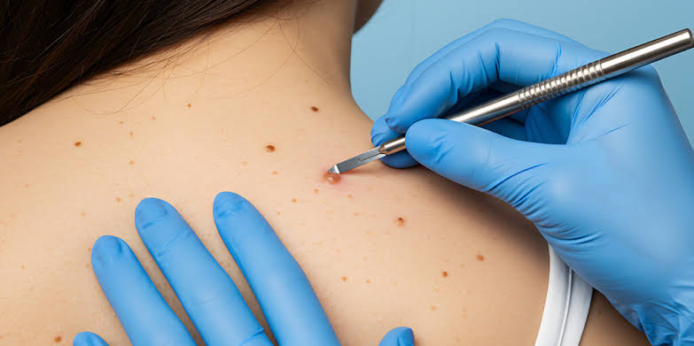
Hearing “we need to biopsy that” tends to trigger more anxiety than the procedure itself deserves. So what happens during a skin biopsy, exactly? It’s one of the quickest, most routine procedures in dermatology, most types take less than 15 minutes, use only local anesthesia, and are done right in the office on the same day your dermatologist spots something worth checking. Knowing exactly what happens during a skin biopsy before, during, and after tends to remove most of the dread.
Why Your Dermatologist Ordered One
A biopsy is the only way to confirm what a lesion actually is. Visual exams and tools like a dermatoscope can raise suspicion, but only a pathologist looking at your tissue under a microscope can tell the difference between a harmless growth and something like basal cell carcinoma, squamous cell carcinoma, or melanoma. If you’re unclear on how your dermatologist decided your spot needed a closer look in the first place, our guide on the types of skin cancer covers what each one tends to look like before it reaches biopsy stage.
The Four Types of Skin Biopsy
Your dermatologist picks the type based on the size, depth, and location of the lesion, and how deep a sample they need to make an accurate diagnosis. According to MedlinePlus, the three most common approaches are shave, punch, and excisional biopsy, with incisional biopsy used less often for larger lesions.
Shave biopsy. The most common type. A blade shaves off a thin sample from the top layers of skin. It’s fast, doesn’t usually need stitches, and works well for raised lesions. It’s generally not used for a lesion your dermatologist suspects could be melanoma, since it may not capture enough depth to measure how far the cells have grown.
Punch biopsy. A small, circular tool is pressed and rotated into the skin to remove a cylindrical sample that goes through multiple layers, including deeper tissue. This is the go-to method when a dermatologist needs full-thickness tissue, for inflammatory skin conditions, deeper pigmented lesions, or when melanoma is suspected and depth matters for diagnosis.
Excisional biopsy. The entire lesion is removed using a scalpel, along with a small margin of surrounding healthy skin. This is typically used when a dermatologist wants the whole growth gone in one step, not just a sample, often for lesions with a higher suspicion of melanoma.
Incisional biopsy. Similar to an excisional biopsy but only removes part of the lesion rather than all of it. This is used when a growth is too large to remove completely in one visit, or sits somewhere that full removal would cause unnecessary scarring or functional problems before a diagnosis is even confirmed.
What Happens During a Skin Biopsy, Step by Step
- The area is cleaned. Your dermatologist disinfects the site and the skin around it to reduce infection risk.
- You get a local anesthetic. A small injection numbs the area completely. This is usually the only part of the procedure you’ll feel, a brief pinch or sting, and it fades within seconds.
- The sample is taken. Depending on the type, this means a blade shaving off a thin layer, a punch tool rotating through the skin, or a scalpel removing all or part of the lesion. You shouldn’t feel pain here, though you may notice pressure or tugging.
- Bleeding is controlled. For shallow shave biopsies, this is often done with a topical solution or light cautery. Punch, excisional, and incisional biopsies that go deeper usually need one or more stitches to close the site.
- The wound is dressed. A bandage is applied, along with instructions on how to care for it over the following days.
- The sample is sent to a lab. Your tissue goes to a pathologist, who examines it under a microscope to determine exactly what it is.
Most biopsies, including consultation and prep time, take under 30 minutes start to finish. Excisional and incisional biopsies that require more extensive cutting and stitching can run closer to 20 to 45 minutes.
Does It Hurt?
The local anesthetic means what happens during a skin biopsy itself shouldn’t hurt. The injection to numb the area is typically the most uncomfortable moment, and it’s brief. Once the area is numb, most people describe feeling pressure or tugging rather than pain. Some soreness at the site once the anesthetic wears off, usually a few hours later, is normal and generally manageable with over-the-counter pain relief if needed.
Healing: What to Expect by Type
Shave biopsy sites tend to look red, shallow, and raw for the first few days, similar to a minor scrape. A thin scab forms within a day or two and gradually lifts as new skin forms underneath. Most shave sites heal within one to two weeks.
Punch biopsy sites heal in a similar timeframe, generally one to two weeks, though sites that required stitches may take slightly longer and need a follow-up visit to have them removed.
Excisional and incisional biopsy sites take the longest, generally two to four weeks, since they involve a larger wound and stitches. Wounds on the legs and feet tend to heal more slowly than those on the face or arms. GoodRx’s breakdown of skin biopsy healing stages is a useful reference if you want to track what’s normal day by day.
Across all types, keep the area clean, apply a thin layer of petroleum jelly if your dermatologist recommends it, and keep it covered with a fresh bandage until it’s fully healed. Avoid picking at the scab, and follow any activity restrictions your dermatologist gives you, especially for sites near joints or areas that see a lot of movement.
When to Call Your Dermatologist
Most biopsy sites heal without any issue, but contact your dermatologist if you notice:
- Increasing redness, swelling, or warmth around the site days after the procedure, rather than gradual improvement
- Pus or a foul odor coming from the wound
- A wound that reopens after it seemed to be closing
- Fever
- Bleeding that doesn’t stop with light pressure
How Long Results Take
Standard turnaround for skin biopsy results is typically 7 to 10 days, though it can range from about 5 to 14 business days depending on the lab and whether additional tests, like special stains, are needed to confirm the diagnosis. Your dermatologist’s office will usually call or schedule a follow-up to go over what the results mean and, if needed, discuss next steps for treatment.
A biopsy confirming basal cell or squamous cell carcinoma typically leads to a conversation about excision, Mohs surgery, or other treatment options depending on the type, size, and location. If you want to understand the difference between those two outcomes ahead of time, our comparison of basal cell carcinoma vs squamous cell carcinoma breaks down how each is typically treated.
FAQs
Is a skin biopsy painful? The procedure itself generally isn’t painful because the area is numbed first. The anesthetic injection is usually the only uncomfortable moment, and it’s brief. Mild soreness after the numbness wears off is normal and usually manageable with over-the-counter pain relief.
Do all skin biopsies need stitches? No. Shallow shave biopsies typically don’t need stitches. Punch biopsies sometimes do, depending on size. Excisional and incisional biopsies almost always require stitches since they involve a larger, deeper wound.
How soon will I know if it’s cancer? Most results come back within 7 to 10 days, sometimes longer if additional lab tests are needed. Your dermatologist’s office will contact you once results are ready rather than leaving you to follow up on your own.
Will a skin biopsy leave a scar? Some scarring is possible with any biopsy that breaks the skin, though shave and punch biopsies typically leave minimal, faint marks. Excisional and incisional biopsies leave a linear scar since they involve stitches, though your dermatologist will discuss ways to minimize its appearance based on the location.
Can I go back to work the same day? For most shave and punch biopsies, yes. Your dermatologist may recommend avoiding strenuous activity or anything that could stress the wound site for a day or two, especially for biopsies on the hands, feet, or joints.