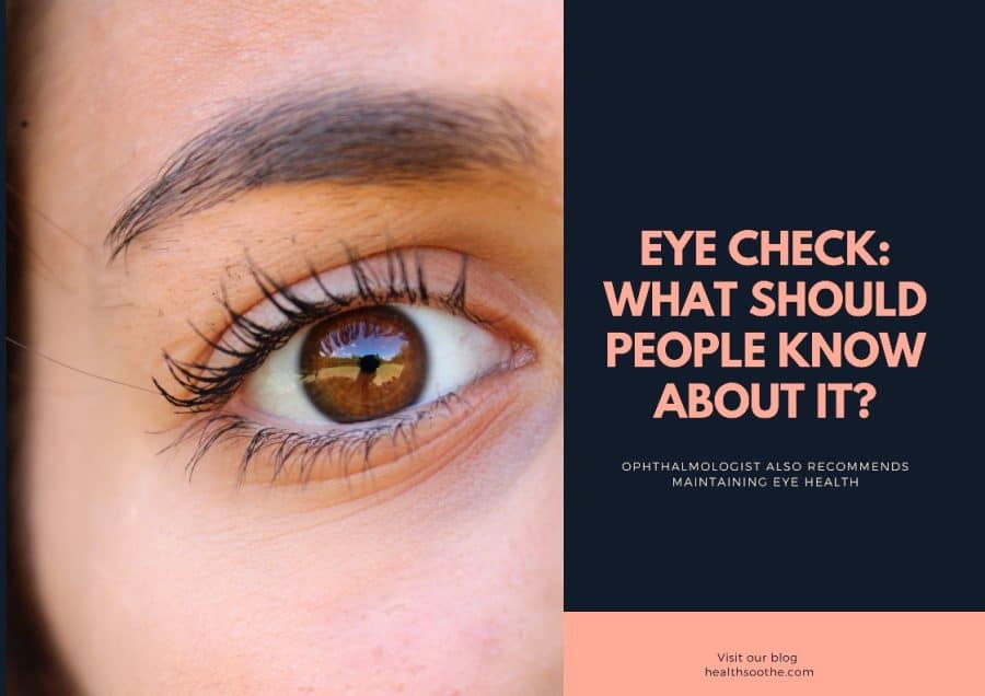
Vision diagnostics is a set of measures for a complete examination of the organs of vision in a specialized eye center or ophthalmological clinic. It is important to understand that not only people with identified vision problems need regular eye examinations but healthy people too. Regular diagnostics are especially important for two groups: children and the elderly.
It is easier to identify pathologies that have just begun to develop in childhood, and they are easier to correct than in adulthood. Children are recommended to conduct a comprehensive vision diagnosis more often than adults: the baby’s vision is regularly checked up to a year, and then, in the period from 1 to 3 years, annually. Mandatory diagnostic periods are the age of 3 and 6-7 years since, at this age, children undergo a medical examination before attending kindergarten/school. It is important to check children's eyesight annually during the school period.
People with vision problems regularly undergo vision diagnostics before correction in optics or the ophthalmologist's office and mistakenly believe that these measures are enough to determine all the problems of the organs of vision. However, in adulthood and old age, it is necessary to undergo visual diagnostics to identify visual impairments, such as cataracts and glaucoma.
A comprehensive examination of the organs of vision is important at any age since many eye diseases are often asymptomatic. The obvious signs may appear in the last stages when treatment requires time, surgery, and hefty costs. But this does not mean that adults must visit an ophthalmologist every month. With no overt symptoms or cause for concern, an eye exam should be done approximately every 6 to 12 months.
How to Prepare for an Eye Exam
Special preparation for the examination of the patient's vision is not required. A few days before the examination, the organs of vision should not be overloaded. The day before the diagnosis of vision, work in the usual mode. On the eve of the examination, it is not recommended to drink alcohol or use psychotropic substances, and do not smoke 30-60 minutes before it.
During the examination, drops will be instilled that dilate the pupil, and other procedures will be performed that can cause tearing and redness of the eyes. Diagnostics can last from 30 minutes to several hours. Therefore, it is important to allow time for dilated pupils to return to normal as visual acuity deteriorates at this point. At this time, it is dangerous to drive, you need to give your eyes a break from gadgets. A patient with dilated pupils should wear sunglasses outside. It is also important not to forget to bring dry disposable handkerchiefs with you. If the patient has the means of vision correction, they should take everything: glasses, lenses with a container, and a solution for washing and storage. Women are not recommended to apply heavy makeup, especially on the eyes and the area around them.
How is Vision Diagnosed?
First, the patient's visual acuity is checked, and the basic diagnostic procedures of the organs of vision are carried out. Next, the patient comes to the ophthalmologist's office, and an initial conversation is held. The doctor learns about the diseases and complaints of the patient and the means of vision correction.
Visual acuity is tested in two ways: by computer diagnostics or using special tables with letters, text, or pictures to test vision.
The main methods of vision diagnostics are:
- determination of visual acuity using various tables for checking vision;
- determination of visual acuity using a digital device (phoropter);
- Schirmer's test - a test for the amount of lacrimal secretions;
- study of color perception - a series of special tests for the sensation and discrimination of colors and their shades;
- autorefractometry - a study of the optical power of the eye using computer diagnostics;
- perimetry - measurement of the visual field of the eye;
- pneumotonometry - non-contact measurement of intraocular pressure;
- echobiometry - determination of the length of the eye;
- biomicroscopy with a narrow pupil - examination of the front of the eye;
- biomicroscopy and ophthalmoscopy with a dilated pupil - examination of the back of the eye and the fundus.
Also, most ophthalmology clinics now use AI in Ophthalmology for OCT (Optical coherence tomography) medical image analysis. This helps patients to be confident that all early, rare diseases will be detected.
After a complete diagnosis, the ophthalmologist conducts the final part of the consultation, during which they decipher and explain to the person all the results. Further, if necessary, the specialist, together with the patient, can choose or change the method of vision correction and choose the treatment. The ophthalmologist also recommends maintaining eye health, working at a computer, etc. If necessary, they set a date for the next consultation or examination at the end of the appointment.
The content is intended to augment, not replace, information provided by your clinician. It is not intended nor implied to be a substitute for professional medical advice. Reading this information does not create or replace a doctor-patient relationship or consultation. If required, please contact your doctor or other health care provider to assist you to interpret any of this information, or in applying the information to your individual needs.

